-
Integrated optical microscope and Laser Scanning Microscope mode
-
Automated sample and tip positioning
-
Soft instruments allow user configurable advanced experiments
-
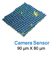
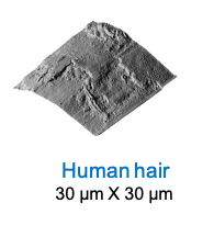
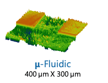
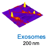
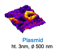
Large feature imaging capability
-
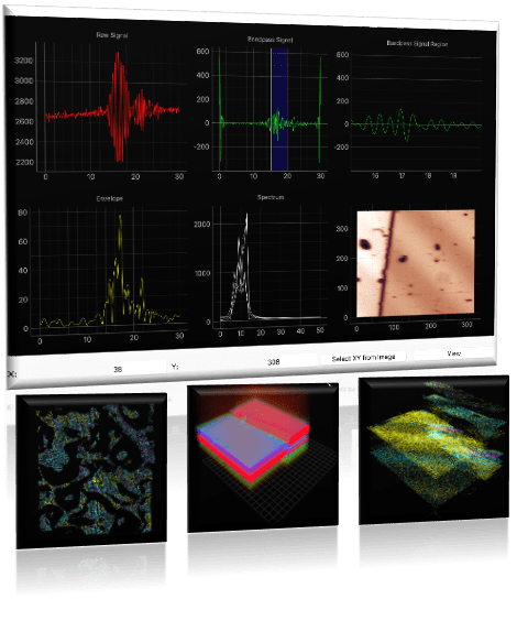
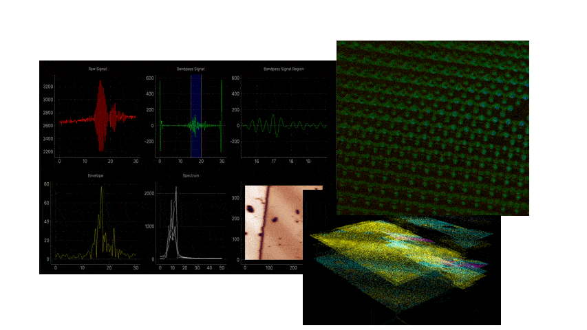
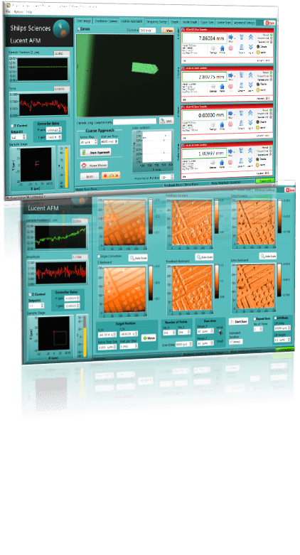
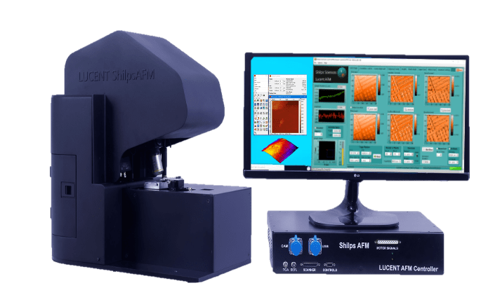
Comprehensive AFM data acquisition software
-
HDF file format saves images, graphs and metadata in one file – compatible with ‘Gwyddion’, the widely used SPM analysis software
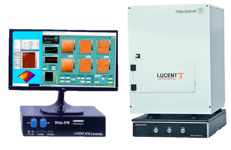
Scanner and positioner
Range (XYZ): 30 µm (Open Loop); 3 µm (Small range setting)
Resolution (XYZ): 0.5 nm (Open Loop); 0.05nm (Small range setting)
Z Noise: 100 pm RMS
Motorized sample (XYZ) and cantilever (Z) positioning: 12 mm; step size< 0.5 µm.
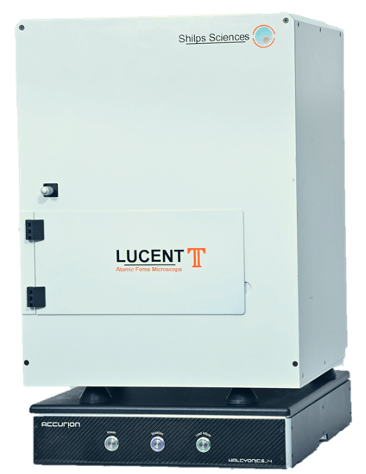
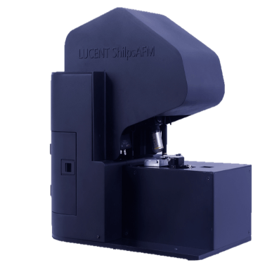
Sample size
Open design allows ample space for a broad class of samples (5 cm x 5 cm x 1 cm).
Electronics and controller
Dedicated FPGA controller with 10kHz loop rate, 19 bits eq AI/AO, Max. image size: 2048 px x 2048 px.
Cantilever sensing
Beam deflection with high sensitivity photo detector,
Typical sensitivity: Force mode – 1 mV/nm, RMS 0.5 mV (150 Hz b/w), Dynamic mode – 10 mV/nm, RMS 2.5 mV (150 Hz b/w)
Optical microscope
10x / 0.3 NA long working distance objective, Working distance 16 mm, 400 µm × 300 µm field of view, Integrated laser & detector for cantilever deflection sensing.
Laser Scanning Microscope
Wavelength & power: 405 nm, ~ 0.5 mW on sample
Spot size: 2.2 µm diffraction limited
Motorized scanning range: 10 mm x 10 mm
Software Features
GUI:
Multiple tabs for easy navigation
AFM Scan settings, Oscillation control, Motors & Camera, LSM scan, Graphing, Advanced.
AFM modes:
Contact mode, Amplitude Modulation, Frequency Modulation, STM.
AFM advanced modes:
User selectable channel for Z-control and an additional channel for data collection.
Conductive AFM, Magnetic Force, Electrical Force.
Shift Mode for point by point Voltage or Z perturbation
User inspired features:
Tip protection, scan direction control, slope correction, multi graphs.
-
Liquid chamber made of inert material for biological studies
-
Electrochemical chamber with electrode connections
-
Soft Instruments module includes sine wave modulation for tip or sample, a low frequency lock-in amplifier (set to f or 2f) and a PI controller
-
STM current amplifier (Sensitivity - 100mV/nA, Range - 100 nA, Bandwidth – 150 Hz)
-
Scanner options - closed loop; Smaller range - 5 µm x 5 µm x 5 µm